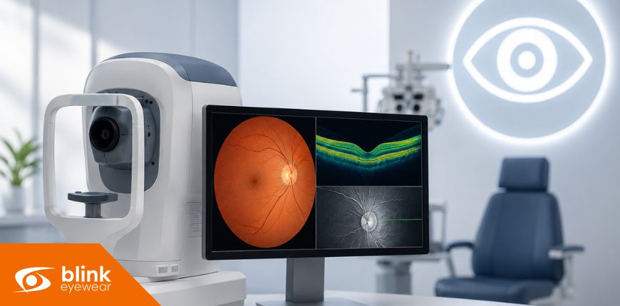
Many eye conditions develop gradually and may not cause noticeable symptoms during their earliest stages. Your vision can seem completely normal even while small changes are occurring in the retina, optic nerve, macula, cornea, or other important structures of the eye. This is one reason comprehensive eye exams are an essential part of protecting your long-term vision.
Modern eye care technology allows optometrists to examine the eyes in greater detail than may be possible with standard vision testing alone. Advanced imaging can produce detailed pictures, measurements, and scans that help identify subtle abnormalities, monitor changes over time, and support more personalized treatment recommendations. These technologies do not replace the knowledge and experience of an eye doctor. Instead, they provide additional information that can help your optometrist.
ADVANCED IMAGING CAN IDENTIFY EYE CONDITIONS BEFORE SYMPTOMS APPEAR
Many people schedule an eye exam because they have noticed blurry vision, headaches, eye strain, or difficulty seeing at night. However, several serious eye conditions can begin without causing obvious changes in everyday vision.
Optical coherence tomography, commonly called OCT, is one of the most valuable imaging technologies used in modern eye care. An OCT scan uses light waves to create detailed cross-sectional images of the retina and optic nerve. These images allow your optometrist to examine the individual layers at the back of the eye and identify changes that may not yet be affecting your sight.
OCT imaging may assist in detecting or monitoring conditions such as:
- Glaucoma
- Macular degeneration
- Diabetic eye disease
- Retinal swelling
- Macular holes
- Retinal membrane changes
- Optic nerve damage
Glaucoma is a good example of why early imaging matters. The condition can gradually damage the optic nerve and reduce peripheral vision. Because these changes may happen slowly, a person may not realize anything is wrong until significant vision has already been affected.
An OCT scan can measure the thickness of the retinal nerve fibre layer surrounding the optic nerve. Your optometrist can compare these measurements with expected ranges and look for areas of thinning that may require further investigation.
Advanced imaging is also valuable for people living with diabetes. High blood sugar can damage the small blood vessels in the retina, potentially causing leakage, bleeding, swelling, or abnormal vessel growth. These changes may not cause immediate symptoms, particularly during the early stages of diabetic eye disease.
Identifying suspicious findings does not always mean that a serious condition is present. Imaging provides your optometrist with additional information that can help determine whether monitoring, further testing, treatment, or a referral to an ophthalmologist is appropriate.
Early detection may allow treatment or monitoring to begin before an eye condition causes permanent vision loss. This makes routine eye examinations important even when your eyesight feels clear and comfortable.
DIGITAL IMAGES CREATE A RECORD FOR MONITORING SMALL CHANGES OVER TIME
A single eye examination provides important information about the current health of your eyes. However, comparing results from multiple appointments can provide an even clearer understanding of how your eyes are changing.
Digital retinal photography captures detailed images of the retina, optic nerve, macula, and blood vessels at the back of the eye. These photographs can become part of your eye health record, allowing your optometrist to compare images from one visit to the next.
This comparison can help reveal whether a freckle, blood vessel, retinal spot, area of pigmentation, or optic nerve feature has remained stable or changed over time. Even small differences may be easier to recognize when current images can be reviewed alongside earlier photographs.
Monitoring changes is especially helpful for patients who have:
- A family history of eye disease
- Diabetes or high blood pressure
- High prescriptions
- Previous eye injuries
- Retinal abnormalities
- Glaucoma risk factors
- Age-related eye changes
For example, the appearance of the optic nerve can vary from person to person. A larger or unusually shaped optic nerve does not automatically indicate glaucoma. By taking images over time, your optometrist can look for progressive changes rather than relying only on a single observation.
Digital retinal photography may also help document retinal freckles, sometimes called choroidal nevi. Many of these spots are harmless, but they may need to be monitored for changes in shape, colour, size, or surrounding tissue.
Advanced imaging creates objective information that can support clinical observations. Instead of relying only on written descriptions, your optometrist can review clear visual records and detailed measurements.
Consistent monitoring is important because not every eye condition requires immediate treatment. In some situations, the most appropriate approach is to observe the area carefully and repeat testing at recommended intervals. Advanced imaging can make this monitoring more accurate and help your eye care provider recognize when additional action may be necessary.
SPECIALIZED MEASUREMENTS SUPPORT BETTER EYE CARE
Different parts of the eye require different types of technology. In addition to retinal photography and OCT scans, optometry clinics may use specialized imaging and measurement tools to examine the cornea, tear film, eye pressure, visual field, or other structures.
Corneal topography creates a detailed map of the curvature and shape of the cornea. The cornea is the clear front surface of the eye and plays an important role in focusing light. Small irregularities in its shape can affect visual clarity and may influence which contact lenses or treatment options are appropriate.
Corneal mapping can be useful when evaluating:
- Astigmatism
- Keratoconus
- Irregular corneal shapes
- Specialty contact lens fittings
- Orthokeratology lenses
- Changes following eye surgery
Patients with keratoconus have a cornea that gradually becomes thinner and more cone-shaped. Detailed corneal measurements can help identify irregularities, monitor progression, and support referrals or treatment planning.
Advanced imaging may also assist with cataract assessment. A cataract develops when the eye’s natural lens becomes cloudy. Although retinal imaging does not directly remove or treat a cataract, a comprehensive examination can help your optometrist evaluate how the cataract is affecting vision and determine whether other eye health concerns are also present.
Symptoms associated with cataracts may include blurry vision, glare, halos around lights, faded colours, reduced contrast, frequent prescription changes, or difficulty driving at night. Your optometrist can combine vision testing, microscope examination, imaging, and your reported symptoms to determine how closely the cataract should be monitored.
For patients experiencing dry eye symptoms, specialized imaging may be used to assess the tear film, eyelids, corneal surface, or oil-producing meibomian glands. This information can help identify possible causes of dryness and guide recommendations based on the individual patient’s needs.
Technology can also support contact lens care. Detailed measurements of the cornea may help your eye doctor select a lens design that provides better comfort, clearer vision, and a healthier fit. This can be particularly valuable for patients with high prescriptions, dry eyes, irregular corneas, or previous difficulties wearing standard contact lenses.
Advanced imaging makes it possible to gather more precise information, but the results must always be interpreted within the context of the complete eye examination. Your symptoms, medical history, family history, medications, prescription, lifestyle, and previous test results all contribute to the final assessment.
Protecting your eyesight involves more than checking whether you can read the smallest letters on a vision chart. A comprehensive eye exam evaluates how well you see while also examining the health of the eye’s internal and external structures.
OCT scans, digital retinal photography, corneal mapping, and other technologies provide valuable information that may help detect concerns earlier, document gradual changes, and guide personalized care. When results are stored and compared over time, your optometrist can build a more complete picture of your long-term eye health.
Regular eye examinations remain important even when you are not experiencing noticeable symptoms. By combining professional expertise with advanced imaging technology, your optometrist can help you understand your eye health, identify potential risks, and take appropriate steps to protect your vision for the future.